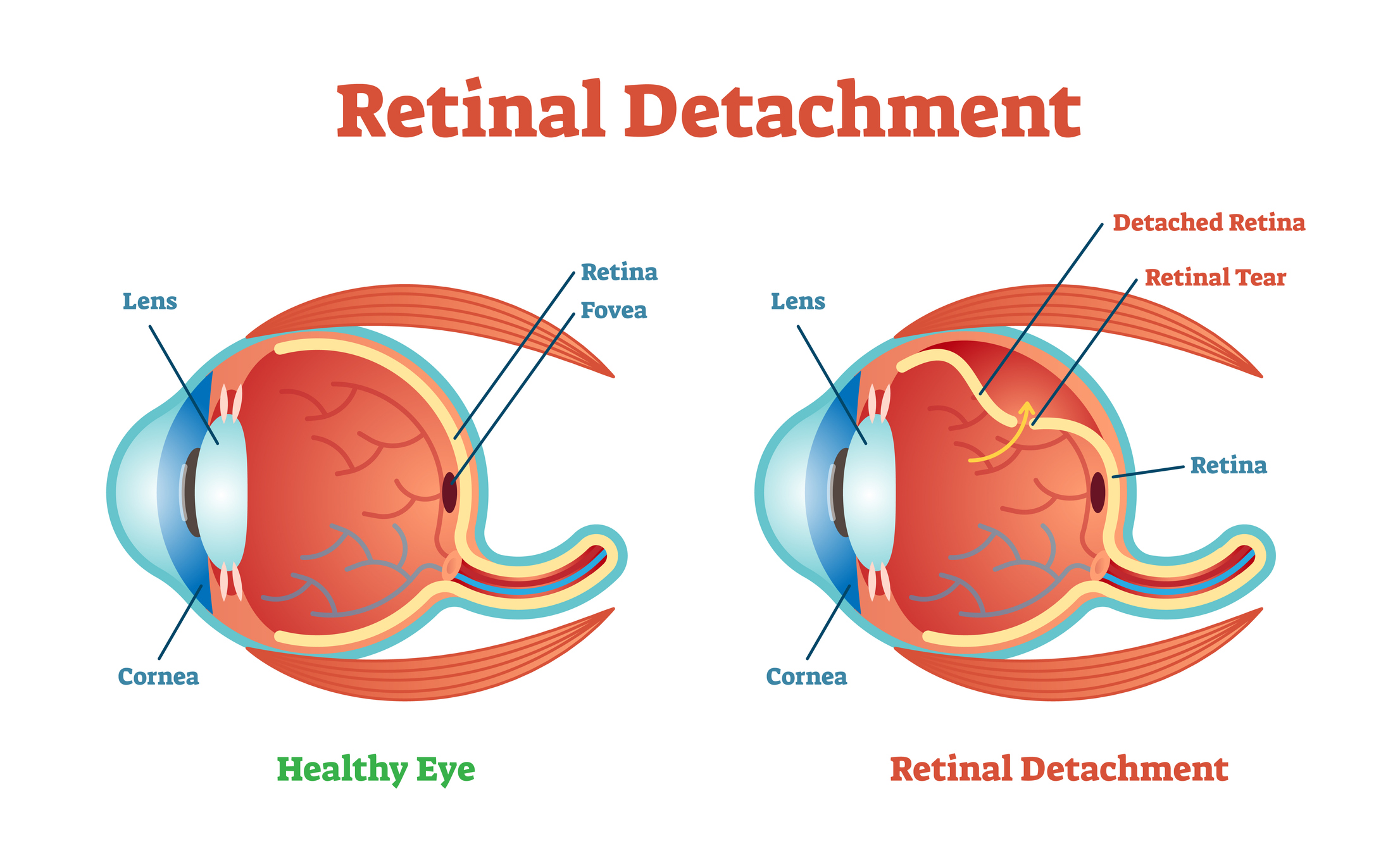
What is a Detached Retina?
Your retina is the layer of nerve cells that line the back wall inside of your eyeball. This specific layer senses light and sends signals to the brain so that you can see. When your retina becomes detached it lifts away from the back of your eye. When your retina is no longer attached to the wall of your eye, it won’t work in the way it’s supposed to therefore your vision will become blurry. This is a serious issue that can cause permanent vision loss if not treated correctly.
How can I tell if I have a Detached Retina?
To diagnose whether or not you may have a Detached Retina, your Doctor Henderson will put drops in your eyes to dilate your pupils. She will then look through a special lens to check to see if anything has changed with your retina.
How would my Retina become Detached?
There is a jelly-like substance that fills the middle of our eyes. This substance is called the vitreous. The vitreous in our eye begins to shrink and become thinner as we grow older. As our eyes move around, the vitreous is supposed to move around on your retina without any issues, but occasionally it can stick to your retina causing painful issues which can lead to tearing the jelly-like substance. If this occurs, the fluid can spill through the tear in your eye and detach your retina.
Who is at risk for a Detached Retina?
The individuals that are more commonly at risk for a detached retina include:
- Those that may need glasses to see far away
- Those who have had cataract surgery
- Suffering from glaucoma
- If you sustain a serious eye injury (harsh blow to the eye)
- Have a retinal tear or detachment in your opposite eye
- Heredity; if your family members have also suffered from retinal detachments
- Have been told by a doctor during an eye exam that you have weak areas in your retinas
What are some of the post-surgery risks?
- Eye infections
- Bleeding in your eye
- Glaucoma if your pressure increases
- Cataracts if it makes your lens become cloudy
- You run the risk of your retina not attaching properly
- You could also run the risk of your retina detaching again
Things you should expect:
- Pain a few hours after surgery
- You need to rest and be less active which means no exercising, no driving, etc.
- Play pirate for a few days! You’ll be wearing an eye patch for however long Doctor Henderson tells you to.
- If a bubble was put in your eye, your head is going to need to stay in that position for about 1-2 weeks. Your ophthalmologist will tell you what position that is and it is very crucial that you follow the instructions exactly
- Floaters and noticing the bubble may be common as your eye is healing a few weeks after surgery.
- Your sight will start to go back to normal about a month post-surgery, however, it could take much longer for it to fully improve and stop changing.

Know the Signs early on
- Flashing lights appear more frequently than usual.
- A detached retina might feel like you’re seeing stars after being hit in the eye.
- Specs, lines, or blurry webs in your field of vision. These are called floaters, and if you noticed an abundance of them at once you could be at risk of a detached retina.
- Shadows appearing in your peripherals
- A curtain covering a portion of your line of vision
How would you treat a Detached Retina?
The only way you can fix a detached retina is surgical. There are three different surgery types to correct this vision issue.
Pneumatic retinopexy:
Doctor Henderson can put a gas bubble inside of your eye. This gas bubble pushes your retina into place so that it can heal properly. After the surgery, you will need to keep your head “face down” for a few days. This will ensure that your gas bubble stays in place and does not move around. As your eye begins to heal, your body will create a fluid that fills your eye. Within enough time, this fluid will take place of where the gas bubble once was.
Vitrectomy:
Doctor Henderson can remove the vitreous membrane by pulling on your retina. She’ll then replace this membrane fluid with either gas or an oil bubble. The bubble pushes into your retina so that it can heal, just like in a pneumatic retinopexy procedure. If she uses the oil bubble method, she can then remove it in a few months. If she fills it with a gas bubble, you won’t be able to fly in an airplane because as the altitude changes, the gas can expand, increase your eyes pressure, and potentially burst.
Scleral buckle:
A band of soft plastic or rubber is sewn to the outside of your eye. This band gently pressures your eye inward. It helps the detached retina to gently heal against the back wall of your eye and is usually left on your eye permanently.
